Last Updated on February 23, 2022
If your horse has a medical problem with his head, your veterinarian may advise that you get a horse skull x ray. If you’ve never seen this process before it can be hard to know what to expect, and you might also be worried that this procedure is painful and unnecessary.
Let’s find out everything you need to know about horse skull radiographs and see what this medical test involves.
Why Would Your Veterinarian Advice A Horse Skull X Ray?
An X-ray, or radiograph, is a diagnostic imaging test used to visualize the inner structures of the horse’s body. This procedure involves pointing a machine very similar to a camera at the area of concern, which fires an X-ray beam through the body. A cassette containing a film is held on the other side of the body, to capture any X-rays that pass through the body.
As the beam passes through the tissues of the body, some are filtered out and others are scattered. The amount that is absorbed depends on the density and type of tissue they pass through. The resulting image on the X-ray film will give your veterinarian a clear picture of the internal structures of your horse’s head.
Whilst we normally associate radiographs with checking for fractured bones, they can also be used to identify other problems. Your veterinarian may advise a horse skull X ray to check for fractures of the skull, or to identify abnormalities of the teeth. Radiographs can also be used to look for problems within the sinuses and oropharyngeal cavity.
Check Out The Ultimate Guide To Different Types Of Ponies
How Are Horse Skull Radiographs Taken?
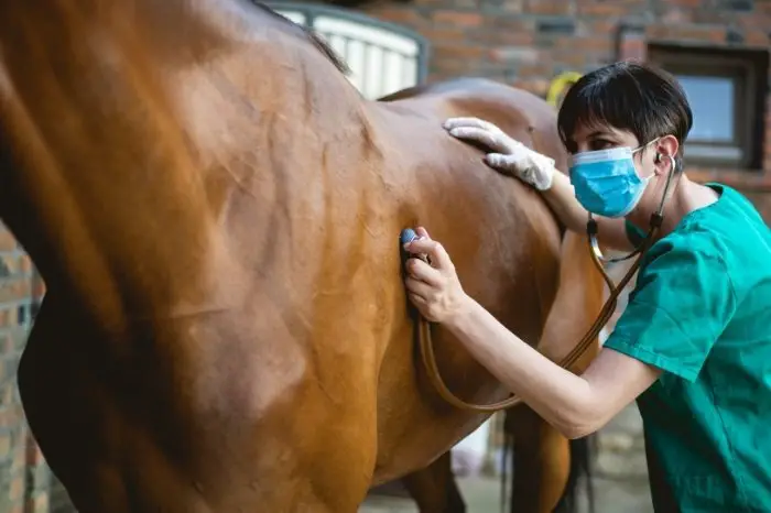
A horse skull radiograph is a non-invasive procedure that will not harm your horse. The veterinary team will want to take a range of radiograph images from different angles, to give a full picture of the horse’s head.
To do this, the horse will need to stand very still for a short period of time. The best way to achieve this is for your veterinarian to administer a small amount of sedative to your horse. The horse will not lie down but will stand very still and not move during the X-ray procedure.
The other reason that sedatives are administered is that it reduces the amount of people necessary to take the radiographs. For veterinary teams who work with radiographs every day, it is essential that they limit their exposure to the X-ray beam as much as possible.
For most medical problems, the X-rays will be taken by holding the film on the side of the head. The X-ray beam may be angled upwards or downwards, to highlight particular structures such as the roots of the teeth.
Formula 707 Calming Equine Supplement – Anxiety Relief and Enhanced Focus for Horses

If your horse has dental problems, your veterinarian may also want to take radiographs of the inside of the mouth. To do this the mouth is held open with a gag, and the X-ray film is placed within the oral cavity.
You may or may not be allowed to be present whilst your horse has his radiographs, depending on the policy of the veterinary center.
Click Here to Learn: Can Horses Have Chocolate?
What Does Equine Skull Radiography Show?
If you have ever seen a radiograph of a horse’s head, you will think that it looks very confusing! It can be difficult to figure out what the different structures are, which is why veterinarians have many years of training to learn to interpret an X-ray film.
However, with a few simple tips, you can figure out what some of the main parts of the horse’s head are on a radiograph. You will see that the X-ray film shows a jumble of white and black shapes, with gray shades in between.
White areas on the radiograph indicate dense tissue, such as bone and tooth. Dark areas are those that are filled with air or fluid, such as the sinuses and oropharyngeal cavity.
The most prominent white area on a horse head X-ray is the cheek teeth – the molars and premolars. You will see two lines of 6 teeth that take up most of the lower part of the skull; one is the upper set of molars, one is the lower set. At the lowest part of the jaw, you will see a set of small teeth called incisors.
If you look above the top row of cheek teeth, you will see a series of large, dark cavities. These are the nasal sinuses, through which your horse breathes. The sinuses connect to the nostrils of the horse at one end, and the oropharynx at the other.
Identifying abnormalities on a radiograph is something that should only be carried out by your veterinarian. They will look for subtle changes on the radiograph, such as fracture lines in the bones or teeth, or pockets of fluid or pus in the nasal sinuses. It may be necessary to take repeat radiographs after treatment to see if any problems have been resolved.
Read more about Horse Canter Vs Gallop – What Is The Difference?
Is An Equine Head X Ray Dangerous?
If you have ever been present when a radiograph was taken, you will see that the medical team take a lot of safety precautions. This is because repeated X-rays can cause long-term health problems.
For an individual person or horse, radiographs are not dangerous. The benefit of a one-off set of radiographs will greatly outweigh the potential risks, and your veterinarian will only advise X-rays if they are absolutely necessary.
You will notice that the veterinary team takes extensive precautions to keep themselves safe during the radiography procedure, using lead gowns and gloves to protect from X-rays.
Summary
So, as we have learned, a horse skull X ray is a diagnostic imaging test carried out by qualified veterinary personnel. This test will give an image of the bones and inner structures of the horse’s head. A horse skull X ray can be used to diagnose medical conditions such as dental disease, sinus infections, and fractured bones.
We’d love to hear your thoughts about equine skull radiography! Has your horse ever had a medical problem diagnosed with a head X ray? Or perhaps you have questions about how a horse skull X ray is carried out? Leave a comment below and we’ll get back to you!
Kate Chalmers is a qualified veterinary nurse who has specialized in horse care for the vast majority of her career. She has been around horses since she was a child, starting out riding ponies and helping out at the local stables before going on to college to study Horse Care & Management. She has backed and trained many horses during her lifetime and competed in various equestrian sports at different levels.
After Kate qualified as a veterinary nurse, she provided nursing care to the patients of a large equine veterinary hospital for many years. She then went on to teach horse care and veterinary nursing at one of the top colleges in the country. This has led to an in-depth knowledge of the care needs of horses and their various medical ailments, as well as a life-long passion for educating horse owners on how to provide the best possible care for their four-legged friends.
Kate Chalmers BSc (Hons) CVN, Dip AVN (Equine) Dip HE CVN EVN VN A1 PGCE
