Last Updated on May 25, 2022
If your horse needs food radiographs, your veterinarian may discuss the benefits of a view of a normal horse foot radiograph versus navicular. Horse foot radiographs have differing levels of complexity, and alternative views are often needed to assess the structures within the hoof walls. Let’s find out everything you need to know about horse foot radiographs!
View Of A Normal Horse Foot Radiograph Versus Navicular
If you need to take your horse for foot radiographs, it can be difficult to know what to expect. Here we will take a look at the different radiographic views used to diagnose common problems of the horse’s foot.
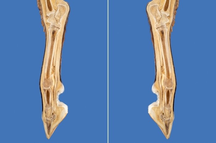
The view of a normal horse foot radiograph versus navicular is used to differentiate between the individual bones in the foot of a horse. The lower limb anatomy of a horse is very complex, and many different radiographic views are needed to assess all the structures accurately. This is one of the most complex areas of a horse to radiograph and requires considerable skill to carry out to a high standard.
Here are some of the most common views that will be taken when a horse has a series of foot radiographs:
Lateromedial Foot Radiographs
The lateromedial view of a horse’s foot is one of the most common types of radiographs you will see performed. This view gives us an image of the horse’s lower limb from the side and is often used to assess horses with laminitis. This is because changes to the position of the distal phalanx can be identified and assessed.
This view also gives a clear image of the joints of the lower limb, helping to diagnosis arthritis and other synovial changes. The navicular bone will be visible on a lateromedial foot radiograph, but not clearly enough to assess it fully.
Dorsopalmar / Dorsoplantar Foot Radiographs
This radiographic view of a horse’s foot is an image taken from the front of the hoof. It is often used to assess the balance of the horse’s hoof, as well as degenerative changes such as ringbone and sidebone. The navicular bone is not clearly visible on this radiographic view, is it is superimposed by the other bones within the hoof walls.
Upright Pedal Radiographs
The upright pedal bone radiographic view is used to give a clear image of the distal phalanx, although it can also be used to assess the navicular bone. Two techniques can be used to acquire this view, with slightly different results.
The first method involves placing the X-ray cassette inside a protective casing on the floor, then standing the hoof of the horse on the cassette. The X-ray machine is placed in front of the horse, with the beam directed downwards through the distal phalanx.
Manna Pro Sho-Hoof Supplement for Horses | Biotin and Zinc Methionine for Healthy Hooves | 5 Pounds

The second technique lifts the hoof so that it is placed flat against the cassette in a special holder. The X-ray beam is directed horizontally to obtain a clear image of the distal phalanx.
With both techniques, the angle of the beam is altered according to the area of interest. It is also necessary to pack any crevices within the foot with putty, as these will show up as artifacts on the final radiograph.
Click Here to Get Info About:
Navicular Skyline Radiographs
This is one of the most challenging radiographic views for an equine radiography technician, as it requires precision and accuracy. The aim is to get a clear radiographic image of the navicular bone, without the superimposition of any other bones of the hoof.
To do this, the X-ray cassette is placed inside a protective holder on the ground, with the horse standing on top of this holder. The leg is positioned so that the hoof is further back than normal, opening up the angle of the pastern. The X-ray machine is placed behind the leg, angled downwards through the navicular bone.
Whilst this is a tricky radiographic view to obtain, it is invaluable in diagnosis problems with the navicular bone in horses. Degenerative changes to the navicular bone are a common cause of lameness in horses, and this radiographic technique is the best way to achieve a clear view of this small but vital bone.

Summary – View Of A Normal Horse Foot Radiograph Versus Navicular
So, as we have learned, the view of a normal horse foot radiograph versus navicular is used to differentiate between the individual bones in the foot of a horse. The lower limb anatomy of a horse is very complex, and many different radiographic views are needed to assess all the structures accurately. This is one of the most complex areas of a horse to radiograph, and requires considerable skill to carry out to a high standard.
We’d love to hear your thoughts on view of a normal horse foot radiograph versus navicular! Have you ever seen a horse having a series of foot radiographs? Perhaps you’ve got some questions about why different radiographs of the horse’s hoof are needed? Leave a comment below and we’ll get back to you!
FAQ’s

Kate Chalmers is a qualified veterinary nurse who has specialized in horse care for the vast majority of her career. She has been around horses since she was a child, starting out riding ponies and helping out at the local stables before going on to college to study Horse Care & Management. She has backed and trained many horses during her lifetime and competed in various equestrian sports at different levels.
After Kate qualified as a veterinary nurse, she provided nursing care to the patients of a large equine veterinary hospital for many years. She then went on to teach horse care and veterinary nursing at one of the top colleges in the country. This has led to an in-depth knowledge of the care needs of horses and their various medical ailments, as well as a life-long passion for educating horse owners on how to provide the best possible care for their four-legged friends.
Kate Chalmers BSc (Hons) CVN, Dip AVN (Equine) Dip HE CVN EVN VN A1 PGCE
